
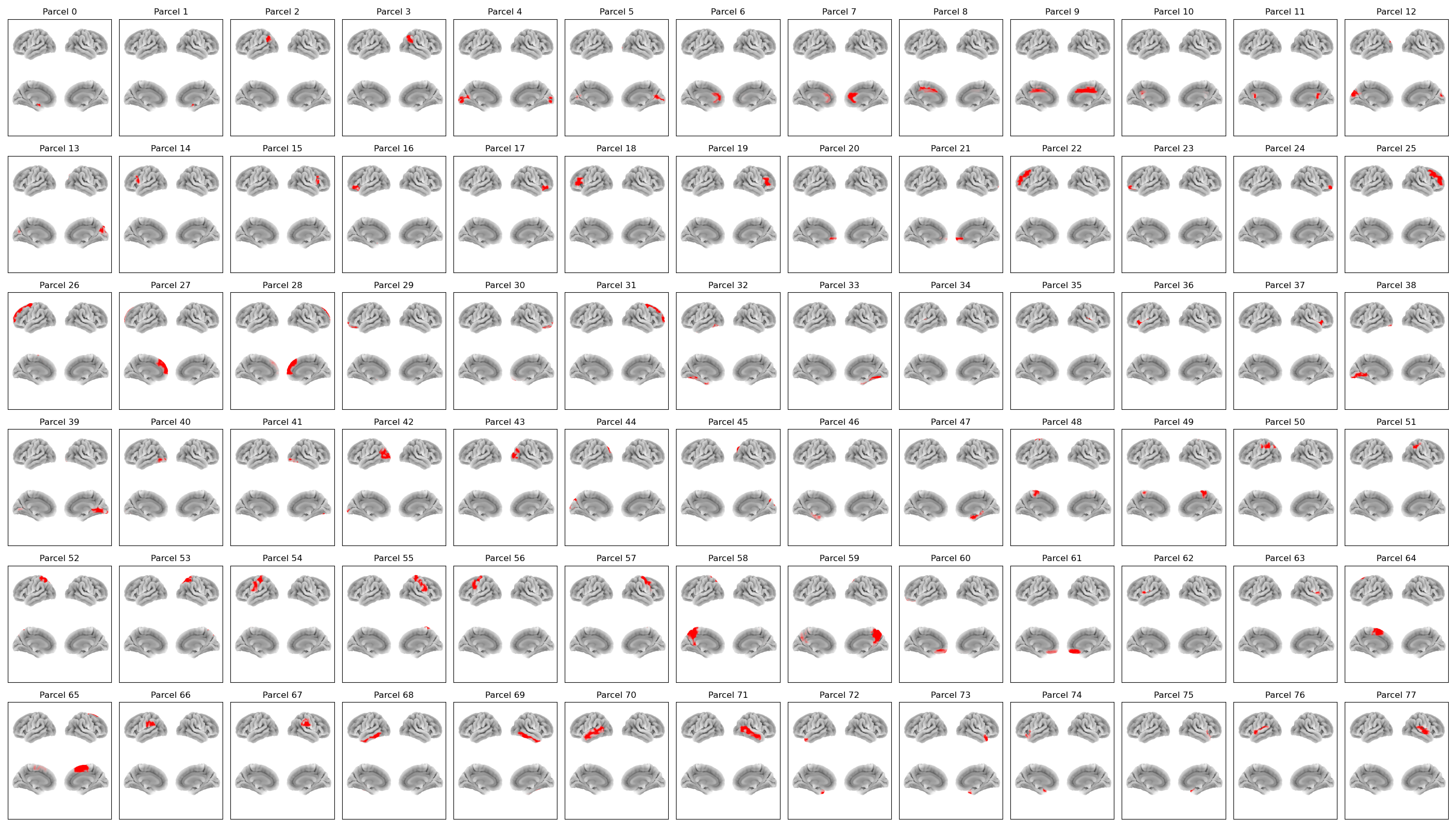
AAL78 Parcellation#
In osl-dynamics, this parcellation file is named atlas-AAL_nparc-78_space-MNI_res-8x8x8.nii.gz, however, this parcellation file was previously named aal_cortical_merged_8mm_stacked.nii.gz (both names will work).
This parcellation contains the cortical regions from the Automated Anatomical Labelling atlas.
Parcels#
Labels and MNI coordinates:
Index |
Parcel |
Hemisphere |
X |
Y |
Z |
|---|---|---|---|---|---|
0 |
Calcarine |
left |
-21.3 |
-1.3 |
-18.7 |
1 |
Calcarine |
right |
28.0 |
0.0 |
-20.0 |
2 |
Occipital_Mid |
left |
-45.3 |
-60.4 |
32.9 |
3 |
Occipital_Mid |
right |
45.4 |
-60.6 |
36.3 |
4 |
Lingual |
midline |
-5.7 |
-80.9 |
4.9 |
5 |
Lingual |
right |
16.0 |
-73.7 |
9.1 |
6 |
Frontal_Sup_Medial |
left |
-5.3 |
35.6 |
11.1 |
7 |
Frontal_Sup_Medial |
right |
7.0 |
36.0 |
13.3 |
8 |
Postcentral |
left |
-7.2 |
-14.8 |
39.5 |
9 |
Postcentral |
right |
7.2 |
-10.5 |
37.9 |
10 |
Precuneus |
left |
-4.0 |
-41.1 |
24.0 |
11 |
Precuneus |
right |
7.4 |
-42.3 |
17.1 |
12 |
Fusiform |
left |
-6.1 |
-82.1 |
24.0 |
13 |
Fusiform |
right |
13.1 |
-81.8 |
25.1 |
14 |
Frontal_Inf_Oper |
left |
-48.8 |
12.4 |
20.4 |
15 |
Frontal_Inf_Oper |
right |
50.1 |
13.2 |
17.4 |
16 |
Frontal_Mid |
left |
-35.7 |
28.6 |
-13.8 |
17 |
Frontal_Mid |
right |
41.1 |
29.4 |
-14.1 |
18 |
Frontal_Inf_Orb |
left |
-45.4 |
28.1 |
11.4 |
19 |
Frontal_Inf_Orb |
right |
50.0 |
28.7 |
12.2 |
20 |
Frontal_Med_Orb |
left |
-6.7 |
49.3 |
-8.0 |
21 |
Frontal_Med_Orb |
right |
7.6 |
53.3 |
-8.9 |
22 |
Parietal_Sup |
left |
-34.0 |
32.5 |
33.5 |
23 |
Temporal_Pole_Mid |
left |
-29.6 |
48.5 |
-12.3 |
24 |
Temporal_Pole_Mid |
right |
34.4 |
51.2 |
-12.8 |
25 |
Temporal_Mid |
right |
38.0 |
32.2 |
32.3 |
26 |
Precuneus |
left |
-18.3 |
32.5 |
42.0 |
27 |
Paracentral_Lobule |
left |
-6.1 |
49.4 |
26.3 |
28 |
Paracentral_Lobule |
right |
8.4 |
51.0 |
27.2 |
29 |
OFCmed |
left |
-15.1 |
47.4 |
-16.6 |
30 |
OFCmed |
right |
16.9 |
42.2 |
-18.7 |
31 |
Temporal_Mid |
right |
22.3 |
30.5 |
41.6 |
32 |
Temporal_Inf |
left |
-32.3 |
-41.4 |
-21.9 |
33 |
Temporal_Inf |
right |
34.6 |
-40.7 |
-21.5 |
34 |
Temporal_Sup |
left |
-44.0 |
-18.7 |
8.0 |
35 |
Temporal_Sup |
right |
44.0 |
-20.0 |
10.7 |
36 |
Insula |
left |
-35.5 |
7.7 |
0.8 |
37 |
Insula |
right |
38.5 |
7.3 |
1.1 |
38 |
Occipital_Inf |
left |
-14.9 |
-71.1 |
-8.0 |
39 |
Occipital_Inf |
right |
15.6 |
-69.5 |
-4.2 |
40 |
Fusiform |
left |
-35.5 |
-81.1 |
-9.6 |
41 |
Fusiform |
right |
37.1 |
-81.6 |
-11.2 |
42 |
Precuneus |
left |
-31.9 |
-82.8 |
14.1 |
43 |
Precuneus |
right |
37.1 |
-81.9 |
16.4 |
44 |
Lingual |
left |
-15.8 |
-85.0 |
27.8 |
45 |
Lingual |
right |
25.2 |
-82.4 |
31.5 |
46 |
ParaHippocampal |
left |
-22.7 |
-16.5 |
-23.5 |
47 |
ParaHippocampal |
right |
25.2 |
-15.2 |
-22.4 |
48 |
Supp_Motor_Area |
left |
-7.8 |
-25.1 |
70.5 |
49 |
Supp_Motor_Area |
right |
7.5 |
-32.5 |
67.5 |
50 |
Angular |
left |
-42.9 |
-49.3 |
45.5 |
51 |
Angular |
right |
48.9 |
-48.0 |
48.3 |
52 |
Precuneus |
left |
-24.1 |
-60.6 |
57.5 |
53 |
Precuneus |
right |
26.3 |
-61.1 |
58.9 |
54 |
Temporal_Mid |
left |
-41.3 |
-25.1 |
48.4 |
55 |
Temporal_Mid |
right |
40.3 |
-27.3 |
51.6 |
56 |
Temporal_Pole_Mid |
left |
-38.8 |
-6.9 |
49.2 |
57 |
Temporal_Pole_Mid |
right |
41.6 |
-8.9 |
50.1 |
58 |
Cingulate_Post |
left |
-8.1 |
-56.7 |
47.3 |
59 |
Cingulate_Post |
right |
9.5 |
-59.1 |
42.7 |
60 |
ParaHippocampal |
left |
-6.4 |
37.6 |
-18.8 |
61 |
ParaHippocampal |
right |
7.3 |
34.8 |
-18.8 |
62 |
Frontal_Inf_Oper |
left |
-47.1 |
-9.8 |
11.7 |
63 |
Frontal_Inf_Oper |
right |
54.4 |
-4.7 |
11.3 |
64 |
Frontal_Sup_Medial |
left |
-5.8 |
5.2 |
59.1 |
65 |
Frontal_Sup_Medial |
right |
8.3 |
0.2 |
59.4 |
66 |
Parietal_Inf |
left |
-55.5 |
-35.1 |
27.5 |
67 |
Parietal_Inf |
right |
56.9 |
-31.5 |
32.0 |
68 |
Temporal_Inf |
left |
-50.2 |
-32.7 |
-22.9 |
69 |
Temporal_Inf |
right |
53.1 |
-32.8 |
-23.2 |
70 |
Fusiform |
left |
-56.8 |
-32.5 |
-4.7 |
71 |
Fusiform |
right |
57.3 |
-39.0 |
-2.7 |
72 |
Temporal_Pole_Sup |
left |
-36.7 |
12.7 |
-36.0 |
73 |
Temporal_Pole_Sup |
right |
45.8 |
12.4 |
-32.4 |
74 |
Temporal_Mid |
left |
-39.3 |
13.8 |
-22.2 |
75 |
Temporal_Mid |
right |
47.0 |
14.5 |
-19.0 |
76 |
OFClat |
left |
-52.0 |
-19.9 |
3.9 |
77 |
OFClat |
right |
57.9 |
-20.6 |
4.3 |
Example Code#
Example code for plotting with this parcellation:
from osl_dynamics.analysis import power

power.save(
    ...,
    mask_file="MNI152_T1_8mm_brain.nii.gz",
    parcellation_file="atlas-AAL_nparc-78_space-MNI_res-8x8x8.nii.gz",
    filename="map_.png",
)
Reference#
If you use this parcellation, please cite:
Tzourio-Mazoyer, N., Landeau, B., Papathanassiou, D., Crivello, F., Etard, O., Delcroix, N., Mazoyer, B., & Joliot, M. (2002). Automated Anatomical Labeling of Activations in SPM Using a Macroscopic Anatomical Parcellation of the MNI MRI Single-Subject Brain. NeuroImage, 15(1), 273-289. https://doi.org/10.1006/nimg.2001.0978