
AAL24 Parcellation#
In osl-dynamics, this parcellation file is named atlas-AAL_nparc-24_space-MNI_res-8x8x8.nii.gz.
This is a reduced version of the AAL116 parcellation, obtained by merging the original Automated Anatomical Labelling regions into 24 broad anatomical groups (10 bilateral pairs plus 4 midline regions).
Parcels#
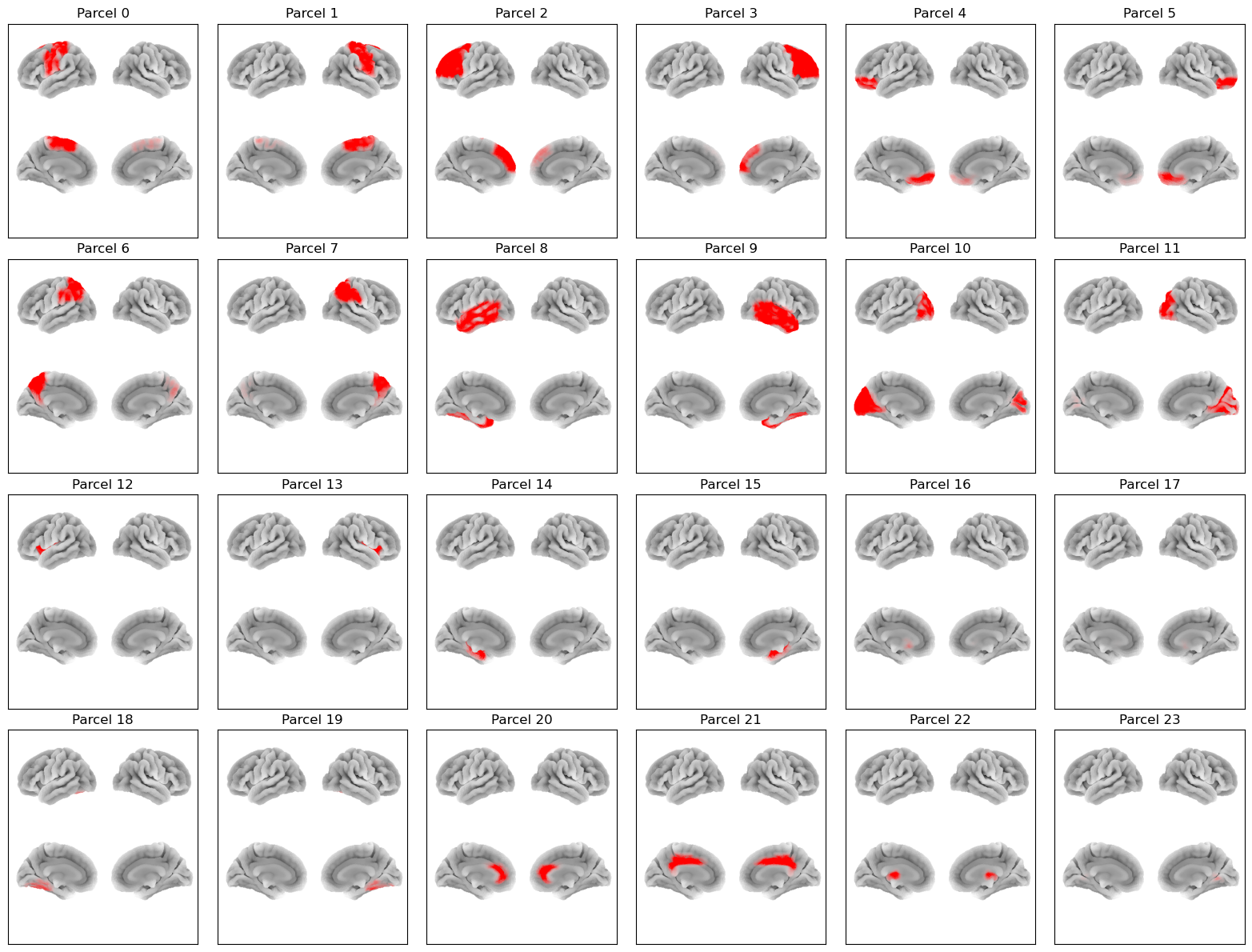
Labels and MNI coordinates:
Index |
Parcel |
Hemisphere |
X |
Y |
Z |
|---|---|---|---|---|---|
0 |
Sensorimotor |
left |
-31.3 |
-13.0 |
50.0 |
1 |
Sensorimotor |
right |
33.2 |
-14.9 |
49.9 |
2 |
Lateral Frontal |
left |
-27.6 |
33.4 |
30.2 |
3 |
Lateral Frontal |
right |
32.0 |
31.6 |
31.0 |
4 |
Orbitofrontal |
left |
-21.5 |
39.0 |
-13.4 |
5 |
Orbitofrontal |
right |
24.5 |
40.1 |
-13.3 |
6 |
Parietal |
left |
-28.9 |
-53.5 |
45.1 |
7 |
Parietal |
right |
32.6 |
-52.8 |
44.4 |
8 |
Lateral Temporal |
left |
-48.0 |
-25.9 |
-12.3 |
9 |
Lateral Temporal |
right |
51.0 |
-26.1 |
-11.9 |
10 |
Occipital |
left |
-19.4 |
-79.2 |
10.0 |
11 |
Occipital |
right |
23.1 |
-77.2 |
11.1 |
12 |
Insula |
left |
-35.4 |
5.5 |
2.2 |
13 |
Insula |
right |
38.7 |
5.1 |
0.8 |
14 |
Medial Temporal |
left |
-23.4 |
-17.6 |
-17.1 |
15 |
Medial Temporal |
right |
26.9 |
-16.5 |
-17.3 |
16 |
Basal Ganglia |
left |
-18.1 |
5.1 |
3.9 |
17 |
Basal Ganglia |
right |
21.2 |
6.2 |
3.9 |
18 |
Cerebellum (lateral) |
left |
-25.5 |
-60.9 |
-35.1 |
19 |
Cerebellum (lateral) |
right |
28.4 |
-60.9 |
-37.1 |
20 |
Anterior Cingulate |
midline |
1.8 |
34.9 |
13.7 |
21 |
Middle Cingulate |
midline |
1.0 |
-18.0 |
36.5 |
22 |
Thalamus |
midline |
0.5 |
-18.8 |
6.8 |
23 |
Cerebellar Vermis |
midline |
1.8 |
-57.8 |
-18.9 |
Each AAL24 parcel was formed by merging the following AAL116 regions:
Parcel |
AAL116 regions |
|---|---|
Sensorimotor |
Precentral, Postcentral, Supp_Motor_Area, Paracentral_Lobule, Rolandic_Oper |
Lateral Frontal |
Frontal_Sup, Frontal_Mid, Frontal_Inf_Oper, Frontal_Inf_Tri, Frontal_Sup_Medial |
Orbitofrontal |
Frontal_Sup_Orb, Frontal_Mid_Orb, Frontal_Inf_Orb, Frontal_Med_Orb, Rectus, Olfactory |
Parietal |
Parietal_Sup, Parietal_Inf, Precuneus, SupraMarginal, Angular |
Lateral Temporal |
Temporal_Sup, Temporal_Mid, Temporal_Inf, Temporal_Pole_Sup, Temporal_Pole_Mid, Fusiform, Heschl |
Occipital |
Occipital_Sup, Occipital_Mid, Occipital_Inf, Calcarine, Cuneus, Lingual |
Insula |
Insula |
Medial Temporal |
Hippocampus, ParaHippocampal, Amygdala |
Basal Ganglia |
Caudate, Putamen, Pallidum |
Cerebellum (lateral) |
Cerebelum_Crus1, Cerebelum_Crus2, Cerebelum_3, Cerebelum_4_5, Cerebelum_6, Cerebelum_7b, Cerebelum_8, Cerebelum_9 |
Anterior Cingulate |
Cingulum_Ant |
Middle Cingulate |
Cingulum_Mid, Cingulum_Post |
Thalamus |
Thalamus |
Cerebellar Vermis |
Vermis_3, Vermis_4_5, Vermis_6, Vermis_7, Vermis_8, Vermis_9, Vermis_10 |
Example Code#
Example code for plotting with this parcellation:
from osl_dynamics.analysis import power

power.save(
    ...,
    mask_file="MNI152_T1_8mm_brain.nii.gz",
    parcellation_file="atlas-AAL_nparc-24_space-MNI_res-8x8x8.nii.gz",
    filename="map_.png",
)
Reference#
If you use this parcellation, please cite:
Tzourio-Mazoyer, N., Landeau, B., Papathanassiou, D., Crivello, F., Etard, O., Delcroix, N., Mazoyer, B., & Joliot, M. (2002). Automated Anatomical Labeling of Activations in SPM Using a Macroscopic Anatomical Parcellation of the MNI MRI Single-Subject Brain. NeuroImage, 15(1), 273-289. https://doi.org/10.1006/nimg.2001.0978